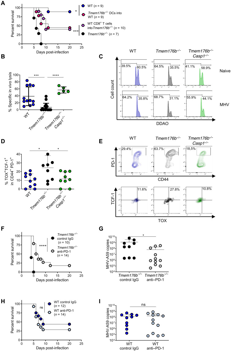
Fig. 2. Inflammasome-dependent T cell exhaustion in Tmem176b−/− mice infected with MHV-A59.
(A) Survival of MHV-A59–infected WT and Tmem176b−/− mice left untreated. In another group, infected WT mice were adoptively transferred with splenic DCs from infected Tmem176b−/− animals, and Tmem176b−/− mice were adoptively transferred with CD8+ T cells from infected WT animals. *P < 0.05; ***P < 0.001, log-rank (Mantel-Cox) test. (B) Percentage of MHV-A59–specific CD8-dependent in vivo lysis was calculated with the formula described in Materials and Methods. Twelve WT, 16 Tmem176b−/−, and 5 Tmem176b−/−Casp1−/− mice were studied at 5 dpi. ***P < 0.001; ****P < 0.0001, one-way ANOVA test. (C) Representative histograms of the experiments shown in (B). (D) Percentage of liver-infiltrating TOX+ TCF-1+ within PD-1+ CD44+ cells, gated in CD8+ MHV-A59–specific T cells. Animals from two independent experiments were analyzed. *P < 0.05, one-way ANOVA test. (E) Representative dot plots of the animals analyzed in (D). (F) Survival of Tmem176b−/− mice infected with MHV-A59 and treated with control IgG or anti–PD-1 antibody. ****P < 0.0001, log-rank (Mantel-Cox) test. (G) Viral load in the liver at 5 dpi with MHV-A59 in Tmem176b−/− mice treated with control IgG or anti–PD-1 antibodies. *P < 0.05, Mann-Whitney test. (H) Survival of WT mice infected with MHV-A59 and treated with control IgG or anti–PD-1 antibody. ns, nonsignificant. Log-rank (Mantel-Cox) test. (I) Viral load in the liver at 5 dpi with MHV-A59 in WT mice treated with control IgG or anti–PD-1 antibodies. Mann-Whitney test.

