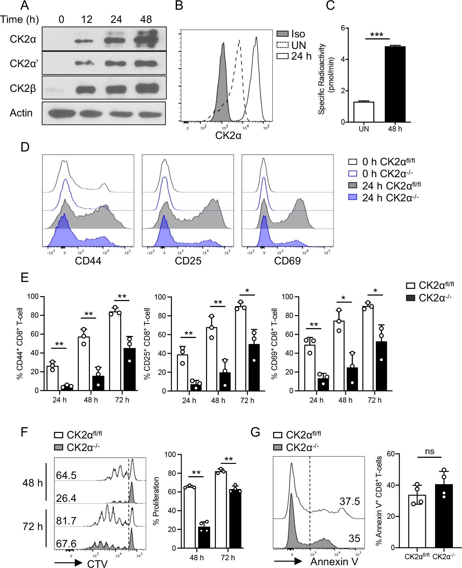
Figure 1. CK2 Subunit Protein Expression and Kinase Activity are Induced in CD8+ T-cells upon Activation, and CK2α is Required for CD8+ T-cell Activation and Proliferation.

Naïve CD8+ T-cells were enriched from the spleen of C57BL/6 mice and activated with plate-bound anti-CD3 (1 μg/ml) and soluble anti-CD28 (1 μg/ml) Abs for the indicated time points. (A) Expression of CK2 subunits CK2α, CK2α’ and CK2β was detected by immunoblotting at the indicated time points. Actin serves as a loading control. (B) CK2α expression was detected by flow cytometry at 24 h under unstimulated conditions (UN) and with anti-CD3 and anti-CD28 Abs treatment. (C) CK2 kinase activity was measured at 48 h by the Casein Kinase 2 Assay Kit. (D) Naïve CD8+ T-cells from CK2αfl/fl and CK2α−/− mice were activated with plate-bound anti-CD3 (1 μg/ml) and soluble anti-CD28 (1 μg/ml) Abs for 24, 48 and 72 h. Expression of CD44, CD25 and CD69 was detected by flow cytometry. Representative line graphs of 24 h are shown. (E) Percentages of CD44, CD25 and CD69 positive cells at the indicated time points are shown. (F) Naïve CD8+ T-cells were labeled with 5 μM CTV, then activated with anti-CD3 (1 μg/ml) and anti-CD28 (1 μg/ml) Abs for 48 and 72 h, and proliferation assessed by CTV dilution. Representative line graphs are shown. (G) Dead and apoptotic cells were assessed by Annexin V staining at 48 h. Representative line graphs are shown. n = 3 in each group. Each experiment was performed two times with three biological replicates per experiment. Bars represent mean ± SD. * p< 0.05, ** p< 0.01, *** p< 0.001. ns = not significant.
