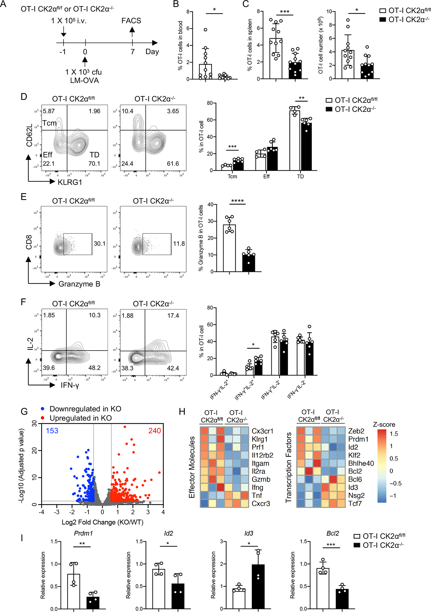
Figure 4. CK2α Promotes CD8+ T-cell Effector Responses during LM-OVA Infection.

(A) One × 105 naïve CD8+ T-cells (CD45.2+) from OT-I CK2αfl/fl or OT-I CK2α−/− mice were adoptively transferred into age- and sex-matched CD45.1 C57BL/6 mice. Twenty-four hour later, recipients were infected with 1 × 10³ cfu of LM-OVA by i.v. injection. Donor CD8+ T-cells from the blood and spleen were analyzed by flow cytometry at day 7. (B) The percentages of OT-I CK2αfl/fl or OT-I CK2α−/− derived CD8+ T-cells in total leukocytes in the blood of recipients at day 7 post-infection are shown. (C) The percentages and numbers of donor CD8+ T-cells in the spleen at day 7 post infection are shown. n=12 in each group. (D) Central memory cells (Tcm), effector cells (Eff) and terminally differentiated effector cells (TD) were detected by CD62L and KLRG1 staining of donor CD8+ T-cells. (E) Granzyme B expression was detected in donor CD8+ T-cells by intracellular staining. (F) IFN-γ and IL-2 expression was detected in donor CD8+ T-cells by intracellular staining. Quantitation of IFN-γ−IL-2+, IFN-γ+IL-2+, IFN-γ+IL-2− and IFN-γ−IL-2− is shown. n = 4–6 in each group. (G) RNA sequencing of OT-I CK2αfl/fl and OT-I CK2α−/− CD8+ T-cells from the spleen at day 7 post infection. Summary of genes differentially regulated by CK2α using the following cutoffs are shown: p < 0.05, fold change >1.5. (H) Heat map shows relative gene expression. (I) Real-time PCR analysis of the indicated genes. n=4 in each group. Each experiment was performed at least two times with two to four biological replicates per experiment, each dot indicates one mouse. Bars represent mean ± SD. * p< 0.05, ** p< 0.01, *** p< 0.001, **** p< 0.0001.
