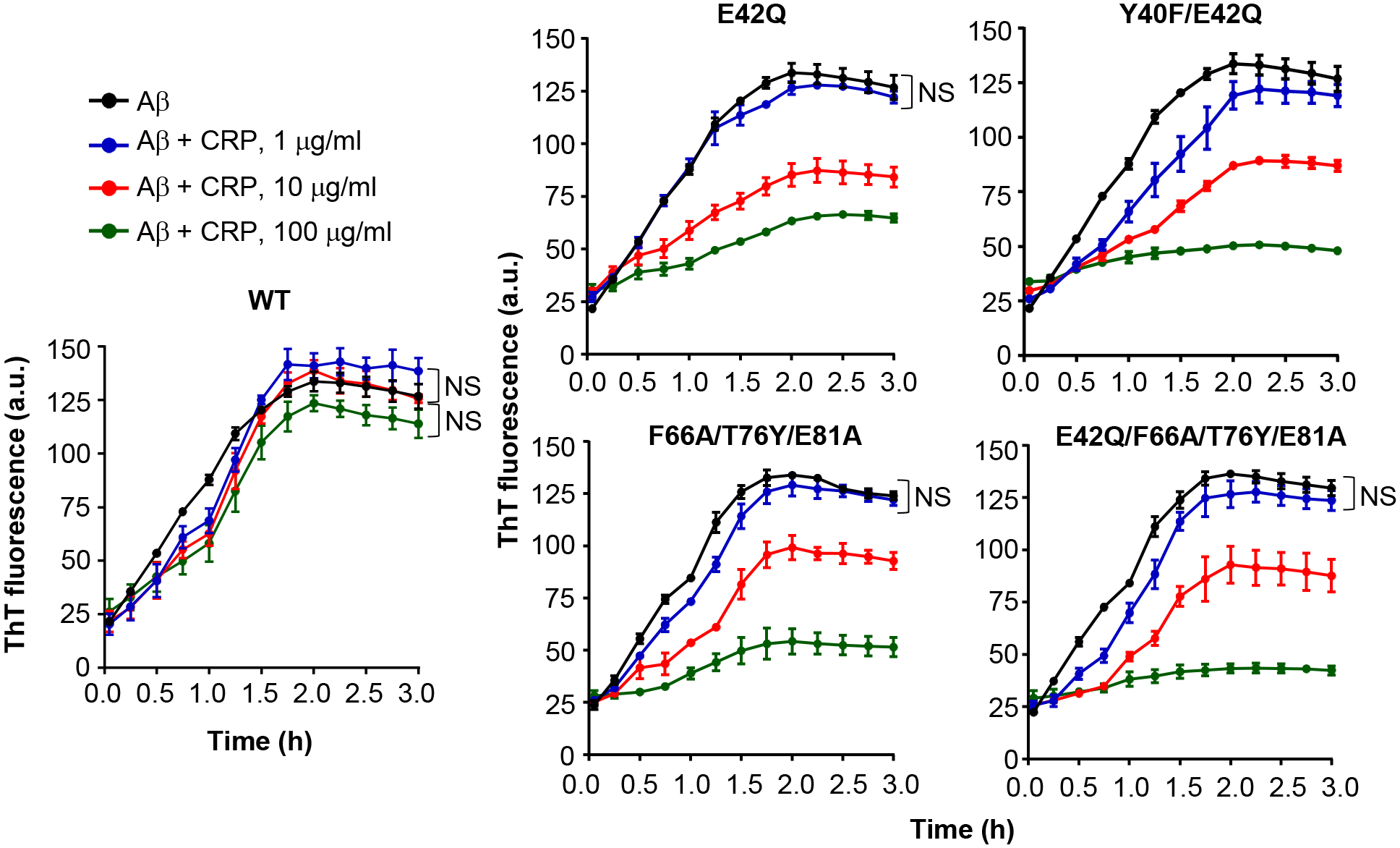
FIGURE 5.

Effects of CRP on the formation of Aβ fibrils. The fibrillation of Aβ peptides was monitored by ThT fluorescence in the absence (black) or presence of 1 μg/ml CRP (blue), 10 μg/ml CRP (red) and 100 μg/ml CRP (green). CRP and Aβ monomers were mixed at time zero and added to microtiter wells. After the first measurement at 5 min, the plate was incubated at 37°C with shaking; fluorescence was measured every 15 min. Results are plotted as mean arbitrary units (a.u.) ± SEM of three experiments. For the time period of 5 min to 2 h, p values were determined by employing linear regression analysis of the slopes. For the time period of 2.25 h to 3 h, p values were determined by taking the mean of all points and employing student unpaired t test. For clarity, only those differences are indicated (NS) where the difference was statistically not significant (p>0.05). The difference between all other curves when compared to each other was statistically significant (p<0.001; not shown).
