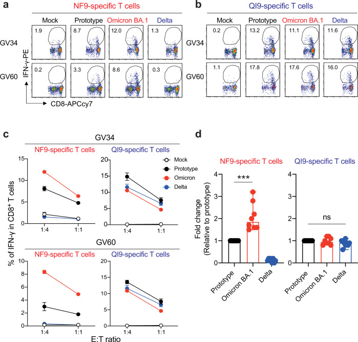
Fig. 2. T-cell recognition to target cells expressing variant spike protein.
a, b HLA-A24-positive T-cell lines of vaccinated donors were stimulated with A549-ACE/A2402 (Supplementary Fig. 1f) expressing spike protein derived from prototype, Omicron, and Delta variants. Representative FACS plots showing intracellular expression of IFN-γ in the NF9-specific CD8+ T cells (a) and QI9-specific CD8+ T cells (b) in two vaccinated donors (GV34 and GV60). c The level of IFN-γ production of NF9- and QI9-specific T cells in response to spike-expressing target cells in two vaccinated donors, GV34 (upper) and GV60 (lower). d Fold changes in IFN-γ expression by NF9-specific T cells (left) and QI9-specific T cells (right) compared to the target cells expressing prototype spike in eight vaccinated donors (GV15, 26, 31, 33, 34, 36, 60, and 61) are shown. a, b The numbers in the FACS plot represent the percentage of IFN-γ+ cells among CD8+ T cells. c The assay was performed in triplicate, and the means are shown with the SD. d The assay was performed in triplicate, and the median is shown. A statistically significant difference versus prototype spike (***p = 0.0002) is determined by a two-tailed Mann–Whitney test. ns, no statistical significance. Source data are provided as a Source Data file.

