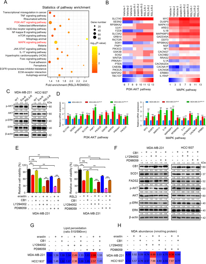
Fig. 5. PI3K-AKT and MAPK pathways were involved in CB1 repressed the ferroptosis sensitivity.
A KEGG pathway analysis of differentially expressed genes in MDA-MB-231 cells transcriptome surviving from increased RSL3 treatment (RSL3-R) (The top 20 most significantly activated pathway are shown). B Heatmap of significantly regulated genes of MDA-MB-231 cells transcriptome surviving from increased erastin treatment (erastin-R) and RSL3 treatment (RSL3-R), respectively, correlated with PI3K-AKT pathway (left) and MAPK pathway (right) (n = 3). C WB analysis of MDA-MB-231 and HCC1937 cells that were resistant to erastin (Era-R) and RSL3(RSL3-R), respectively. D qPCR analysis of the indicated gene expression associated with PI3K-AKT pathway (left) and MAPK pathway (right) in MDA-MB-231 cells that were resistant to erastin (MDA-MB-231Era-R) and RSL3 (MDA-MB-231RSL3-R), respectively, compared with parental MDA-MB-231 cells. **p < 0.01 (t-test). E The cell viability of MDA-MB-231 cells stably transfected with empty vector or CB1 treated with LY294002 (5 μM) or PD98059 (10 μM). The cell viability was assessed by CCK-8 assay after treatment for 72 h with the indicated doses of the drugs. ns (no significance), **p < 0.01 (one-way ANOVA). F WB analysis of MDA-MB-231 and HCC1937 cells transfected with empty vector or CB1. Cells were treated with LY294002 (5 μM) or PD98059 (10 μM). G, H Effect of CB1 overexpression, LY294002 (5 μM) and PD98059 (10 μM) on lipid peroxidation (G) and MDA (H) response to erastin treatment, respectively, in MDA-MB-231 and HCC1937 cells. Data shown are mean ± SD of triplicate measurements that were repeated three times with similar results.

