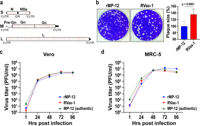
Fig. 1. Genetic structure and growth kinetics of RVax-1 in Vero or MRC-5 cells.
a Schematic representation of the RVax-1 S, M, and L segments, which encode a deletion at nt. 21–384 in the M-segment, and 566 silent mutations throughout the N, M, and L ORFs. Locations of individual silent mutations are labeled in red lines, which are found as a cluster every 50 nt. b Plaque phenotypes of rMP-12 and RVax-1 in Vero cells at 4 dpi. The graph represents the means ± standard deviations of relative diameter lengths from 20 randomly selected plaques. Unpaired t-test was used for the comparison of two groups (t = 7.319, df = 38). Vero cells (c) or MRC-5 cells (d) were infected with MP-12 vaccine lot-7-2-88, rMP-12 or RVax-1 in Vero cells at MOI 0.01. Virus replication kinetics at 35 °C are shown. Means ± standard deviations of triplicate wells are shown.

