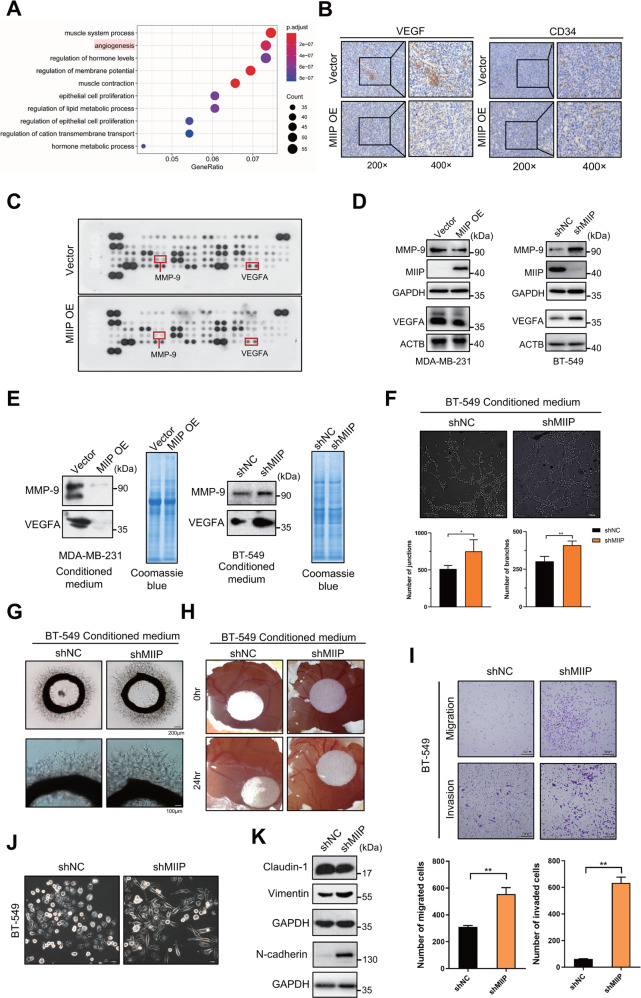
Fig. 3. MIIP inhibits tumor angiogenesis and cell migration of TNBC.
A Gene Ontology analysis of downregulated genes in MIIPhigh expression group relative to MIIPlow expression group using data retrieved from TCGA database. B Expression of CD34 and VEGF in the tumors formed from MDA-MB-231 cell with MIIP overexpression or the control cells were determined by immunohistochemistry. C Human angiogenesis array was performed to determine changes of angiogenesis-related factors in MDA-MB-231 cells after MIIP was overexpressed. D Effect of MIIP on the expression of VEGFA and MMP-9 was validated by western blot in MDA-MB-231 cells and BT-549 cells. E Levels of VEGFA and MMP-9 in the conditioned medium (with same amount in total protein) of MDA-MB-231 cells and BT-549 cells with altered MIIP expression were determined by western blot. Coomassie blue staining of the gel was applied to serve as loading control. F Tube formation assay of HUVEC cells treated with conditioned medium (4 μg in protein amount) from BT-549 cells with altered MIIP expression. Effect of conditioned medium (4 μg in protein amount) from BT-549 cells with or without MIIP knockdown on the ex vivo angiogenesis was evaluated by G rat aortic ring assay and H chick embryo chorioallantoic. I Transwell assay was performed to investigate the abilities of migration and invasion in BT549 cells with indicated stable transfection. J Morphology of BT-549 cells with altered MIIP expression was observed under the bright field of inverted microscope. K Expression levels of EMT-related proteins in BT-549 cells stably transfected with shRNA targeting MIIP (shMIIP) or negative control shRNA (shNC) were determined by western blot. Data are presented as mean values ± SD. *: P < 0.05; **: P < 0.01.

