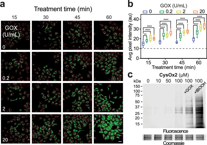
Fig. 4. Cell permeable CysOx probes for no-wash live-cell microscopy of sulfenic acid in cells.
a No-wash live-cell confocal images of HeLa cells at different time points after the addition of CysOx2 (10 µM) and GOX at the indicated concentrations (0–20 U/mL). λex = 458 nm; scale bar: 50 µM. Representative images are shown from N = 3 independent experiments. b Box and whisker plot of the average pixel intensities from panel a. Data are representative of ten independent readings from five different frames. Bars denote ±SEM. Variance was analyzed by two-way ANOVA test. At 15 min: **P = 0.0099 and ****P < 0.0001 when compared against cells treated with probe only. At 30, 45, and 60 min: ****P < 0.0001 when compared against cells treated with probe only. c Representative in-gel fluorescence analysis of lysates derived from HeLa cells incubated with CysOx2 and GOX (20 U/mL) or t-BOOH (500 µM). N = 2 independent experiments. Fluorescence lane intensity is quantified and shown in Supplementary Fig. 9e.

