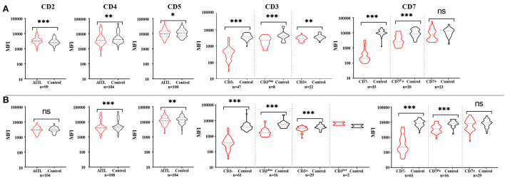
Figure 1.
The expression features of pan-T cell antigens of AITL patients in tissue (A) and bone marrow (B). (A) Compared with the corresponding non-neoplastic CD4+ T cells in AITL, neoplastic T cells had bright CD2 expression and diminished CD3, CD4, and CD5 expression. CD7 mean fluorescent intensity (MFI) had no significant difference between CD7+ neoplastic T cells and normal CD4+ T cells. (B) In bone marrow samples, neoplastic T cells had obviously diminished CD3, CD4, and CD5 expression, and there were no significant difference of CD2 expression between neoplastic T cells and normal CD4+ T cells. CD7 mean fluorescent intensity (MFI) had no significant difference between CD7-positive neoplastic T cells and normal CD4+ T cells. *p < 0.05, **p < 0.01, ***p < 0.001.

