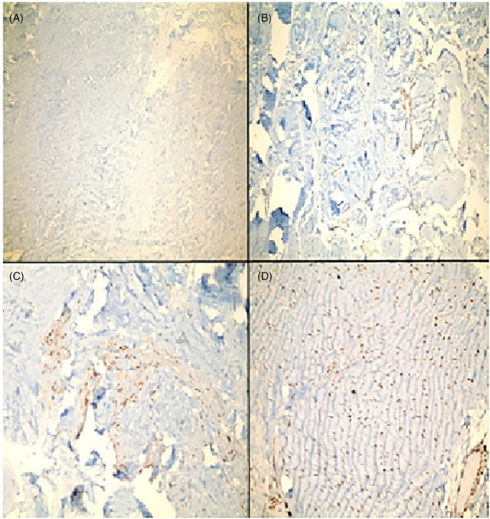
FIGURE 1.

(A‐D) Microscopic images of samples after immunohistochemical staining. (A) TIMP 2 without immune staining, which we accepted as score 0. (B) Immune staining of TIMP 2, less than 10% and accepted as score 1. (C) Immune staining of TIMP 1, between 10% and 50% and accepted as score 2. (D) Immune staining of TIMP 1, over 50% and accepted as score 3. TIMP, tissue inhibitor metalloproteinases
