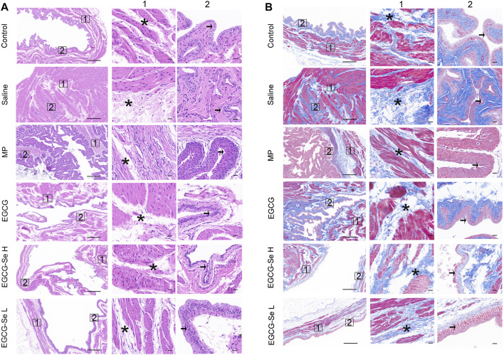
FIGURE 5.
Hematoxylin-eosin (H and E) and Masson staining of bladders. (A) Representative bladders stained with H&E. Columns 1 and 2 are magnifications of the areas marked by the blue squares in the left column. Pathological changes of the bladder are indicated using asterisks (muscle layer) and arrows (intima). Scale bars are 500 and 50 μm, respectively. (B) Representative bladders stained with Masson stain. Columns 1 and 2 are magnifications of the areas marked by the blue squares in the left column. Pathological changes of the bladder are indicated using asterisks (muscle layer) and arrows (intima). Scale bars are 500 and 50 μm, respectively.

