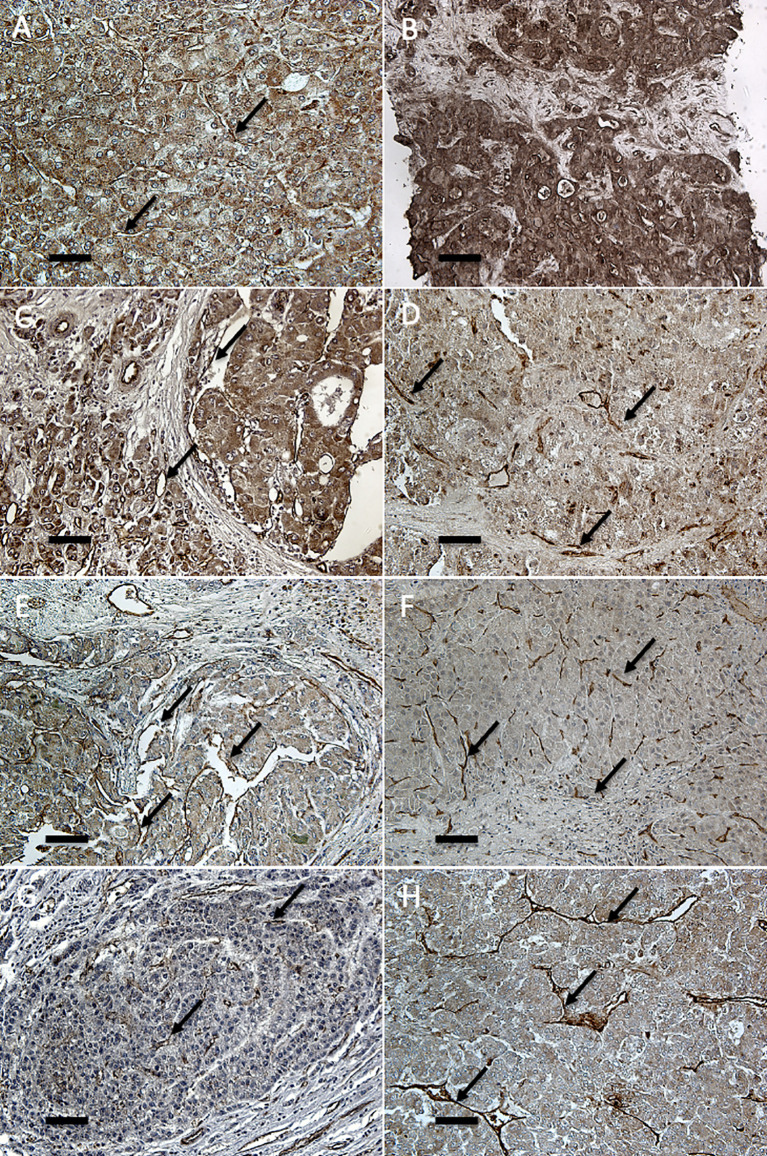
Figure 1.
Immunohistochemical analysis of angiopoietin-2 in endothelia of primary and recurrent HCCs after LT. Immunostaining for angiopoietin-2 (20×) shows marked cytoplasmic and vascular endothelial positivity both in primary HCCs at explant (A, C, E, G) and in the respective recurrent tumors (B: liver; D: peritoneum; F: lung; H: kidney). Endothelial and parenchymal angiopoietin-2 expression in hepatic and extra-hepatic recurrent HCCs is more marked than in the corresponding primary tumor. Arrows indicate representative endothelial localization (scale bar: 6.7 μm).

