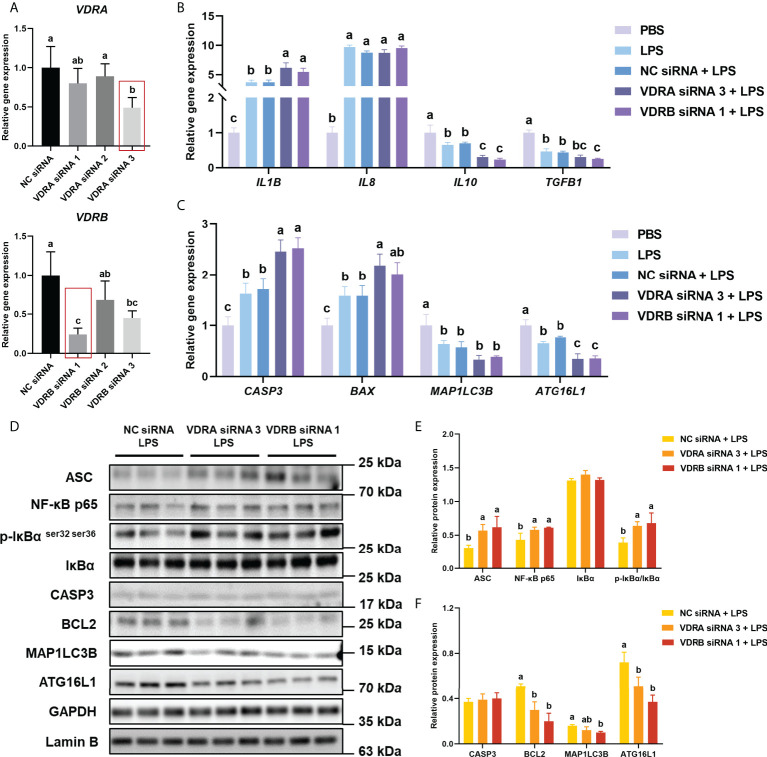
Figure 3.
VDR knockdown in vivo on inflammation, apoptosis, and autophagy in the intestine. (A) The relative mRNA expression level of VDRA&B in the intestine treated with VDRA&B siRNAs. (B) The mRNA expression levels of IL1B, IL8, TGFB1, and IL10. (C) The mRNA expression levels of CASP3, BCL2, BAX, MAP1LC3B, and ATG16L1. (D-F) The level of ASC, intranuclear NF-κB p65, total IκB and phosphorylated IκB, CASP3, BCL2, MAP1LC3B, and ATG16L1 were analyzed and quantitated by western blot. The blots of ASC, IκBα, p-IκBα ser32 ser36, CASP3, BCL2, MAP1LC3B, and ATG16L1 were used for GAPDH loading control, while the blot of NF-κB p65 was used for Lamin B loading control. Error bars of columns denote SD (n = 3), and columns with different letters above them are significantly different (P < 0.05).

