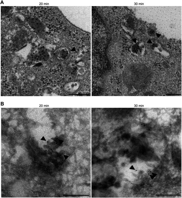
Fig. 5.
EV71 particles are incorporated into LEs. (A) Transmission electron microscopy. RD-hSCARB2 cells were infected with EV71 and fixed at 20 or 30 mai. Times indicate the time after infection. Arrowheads indicate representative EV71-like particles in LE. (B) Immuno-electron microscopy. Gold particles label EV71 antigen. Arrowheads indicate representative gold particles existed in the lumen of single membrane compartments without ILV at 20 mai, and representative gold particles accumulated in single membrane organelles containing ILVs at 30 mai. The experiments were repeated two times and the same trends were observed. Scale bars: 200 nm.

