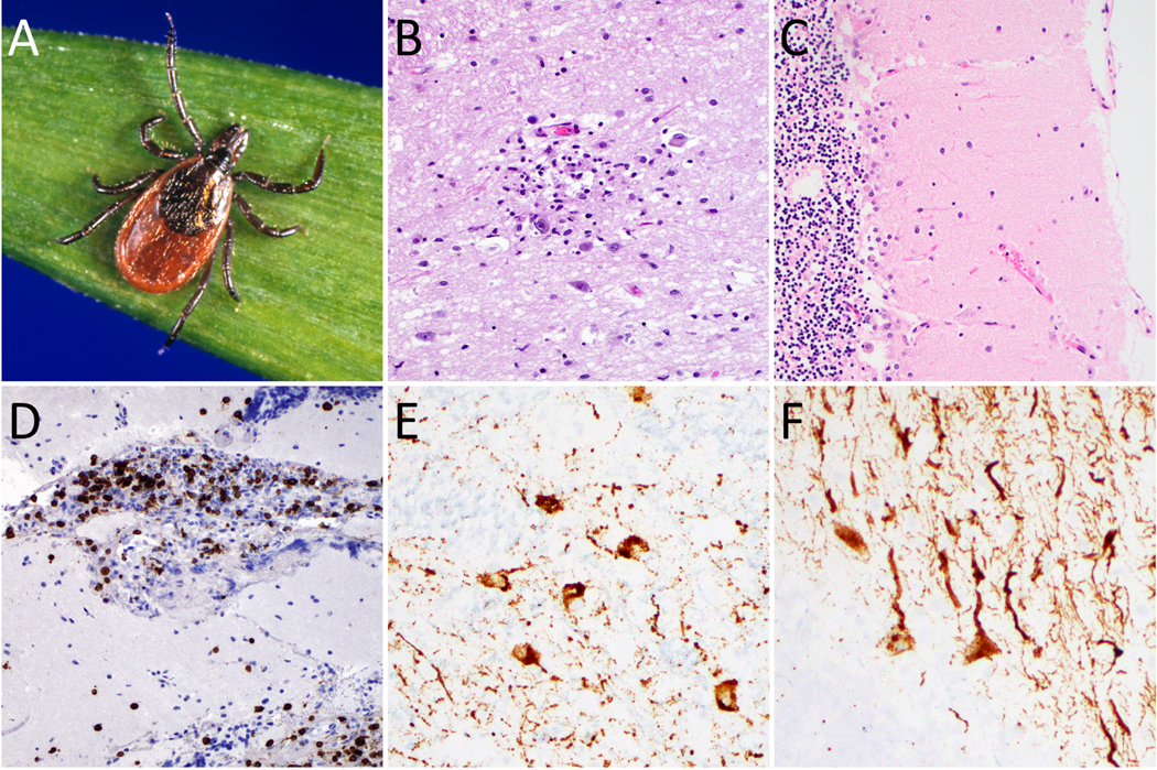
Figure 2: Powassan virus neuropathological features.
Female Ixodes scapularis (deer tick, black-legged tick) can be grossly identified by characteristic U-shaped anal groove (A). Sections from fatal Powassan encephalitis cases show a microglial nodule with neuronophagia in the thalamus (B), marked Purkinje cell loss and Bergmann gliosis in the cerebellum (C), and CD3-positive T cell infiltrate in the leptomeninges (D). Powassan virus RNA in situ hybridization highlights infected neurons in the thalamus (E) and cerebellum (F). (Image A courtesy of Michael L. Levin, PhD via the CDC Public Health Image Library)

