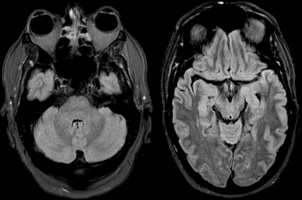
Figure 4: Brain MRI -.
MRI from patient in Box 2 at four days from initial symptom onset shows mild T2/FLAIR hyperintensity in the cerebellar vermis, pons, midbrain, and medial temporal lobes. No significant contrast enhancement or diffusion restriction was identified.

