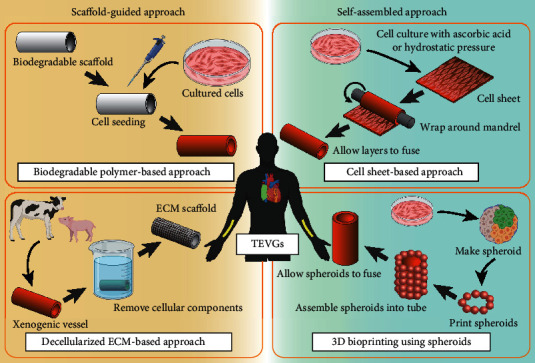
Figure 1.

Schematic representation of TEVG manufacturing methods. Four representative fabrication methods are depicted. TEVGs fabricated as depicted in the right (green) panels are self-assembled approaches, while TEVGs fabricated as depicted in the left (yellow) panels are scaffold-guided approaches. TEVGs fabricated according to these methods are intended as either large-diameter venous shunts in pediatric patients with congenital heart disease (placement shown in green in human schematic) or small-diameter arteriovenous shunts in adult patients with renal disease (placement shown in yellow). This review introduces the spheroid-based technique as a novel application of 3D bioprinting (lower right panel). Graphical objects were created with BioRender (https://biorender.com/).
