Fig. 2.
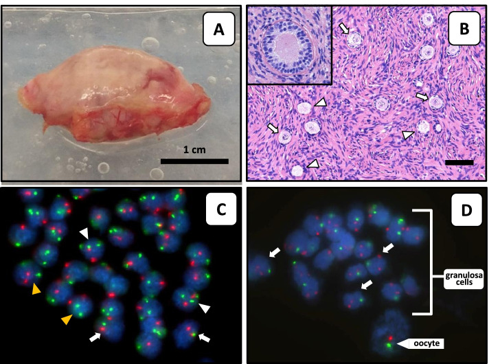
Macroscopic, histologic and FISH analysis of ovarian tissue. A Directly after unilateral ovariectomy the ovary was transported to the laboratory for preparation of cortex fragments for fertility preservation purposes. B Haematoxylin–eosin stained section of cortex tissue with empty follicles (arrows) and follicles with missing granulosa cells (arrow heads). The inset shows a multilaminar follicle with no recognizable oocyte. FISH analysis of cortex stromal cells (C) and of cells from a single follicle (D) with X-chromosome (green) and chromosome 18 (red) specific probes. Arrows point to 45, X cells, white arrow heads to 47, XXX cells and yellow arrow heads to 46, XX cells. The FISH signals from the oocyte are indicated (D). Note that FISH signals are not all in the same focus plane

