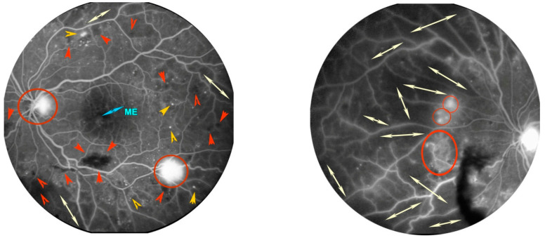
Figure 3.
Fluorescein angiography of a patient with proliferative diabetic retinopathy (DR) in left eye. Posterior pole in the left image. Presence of numerous microaneurysms as hyperfluorescent dots (orange arrow heads), bleeding as irregular black spots (red arrow heads), and initial macular edema (ME) as retinal foveal hyperfluorescence. Retinal nasal quadrant in the right image. Presence of large dark areas without terminal vessels due to retinal ischemia (white arrows). In both images, some hyperfluorescent spots on the optic disc and along the nasal and temporal vascular arches are highlighted, due to proliferation of new blood vessels (red circles).

