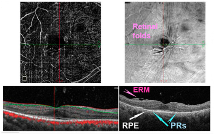
Figure 5.
Optical coherence tomography angiography (OCTA) in a patient with retinitis pigmentosa (RP). In right eye, it is possible to notice the absence of photoreceptors (PRs) (blue arrows) and a retinal pigment epithelium (RPE) degeneration (white arrows) beyond the perifoveal area. The vascular plexus is reduced (upper left image). Moreover, an epiretinal membrane (ERM) is detected in the enface image (upper right image) and structural image (bottom right panel) forming some retinal folds.

