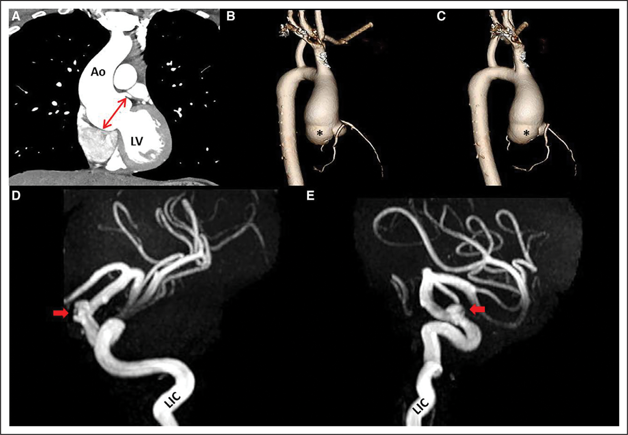
Figure 1. Chest computed tomography angiography and brain magnetic resonance angiography.

A, Chest computed tomography angiogram demonstrates left ventricular (LV) outflow with dilated root (double-headed arrow) and proximal ascending aorta (Ao); 3-dimensional reconstruction shows root phenotype with enlarged sinuses in lateral view (B) and posterior view (C) with proximal ascending involvement. Note disproportional dilatation of noncoronary sinus (asterisk). The remainder of the distal thoracic aorta was normal. Magnetic resonance angiogram of the brain shows left supraclinoid intracranial aneurysm (arrows), originating from the left internal carotid artery (LIC), in lateral (D) and left anterior oblique (E) views.
