
Figure 1. iNKT cell development is severely impaired in T-Mettl14−/− mice.
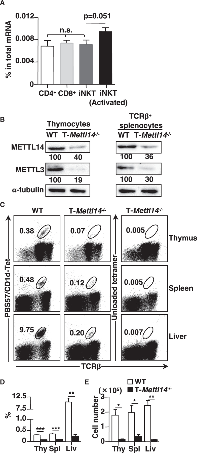
(A) m6A level in total mRNA of CD4+, CD8+ T cells, and iNKT cells (naive and activated) (n = 5–6).
(B) Immunoblot of METTL14 and METTL3 in total thymocytes and TCRβ+ splenocytes in WT and T-Mettl14−/− mice. Data representative of three independent experiments.
(C) Representative staining of lymphocytes in indicated organs from WT and T-Mettl14−/− mice with CD1d/PBS57 tetramer or unloaded CD1d tetramer.
(D and E) Summary of frequencies and cell numbers of iNKT cells in the indicated organs from WT and T-Mettl14−/− mice (n = 5–8). SEM is shown. *p < 0.05, **p < 0.01, ***p < 0.001.