
Figure 3. Mettl14 deficiency impairs the maturation of iNKT cells.
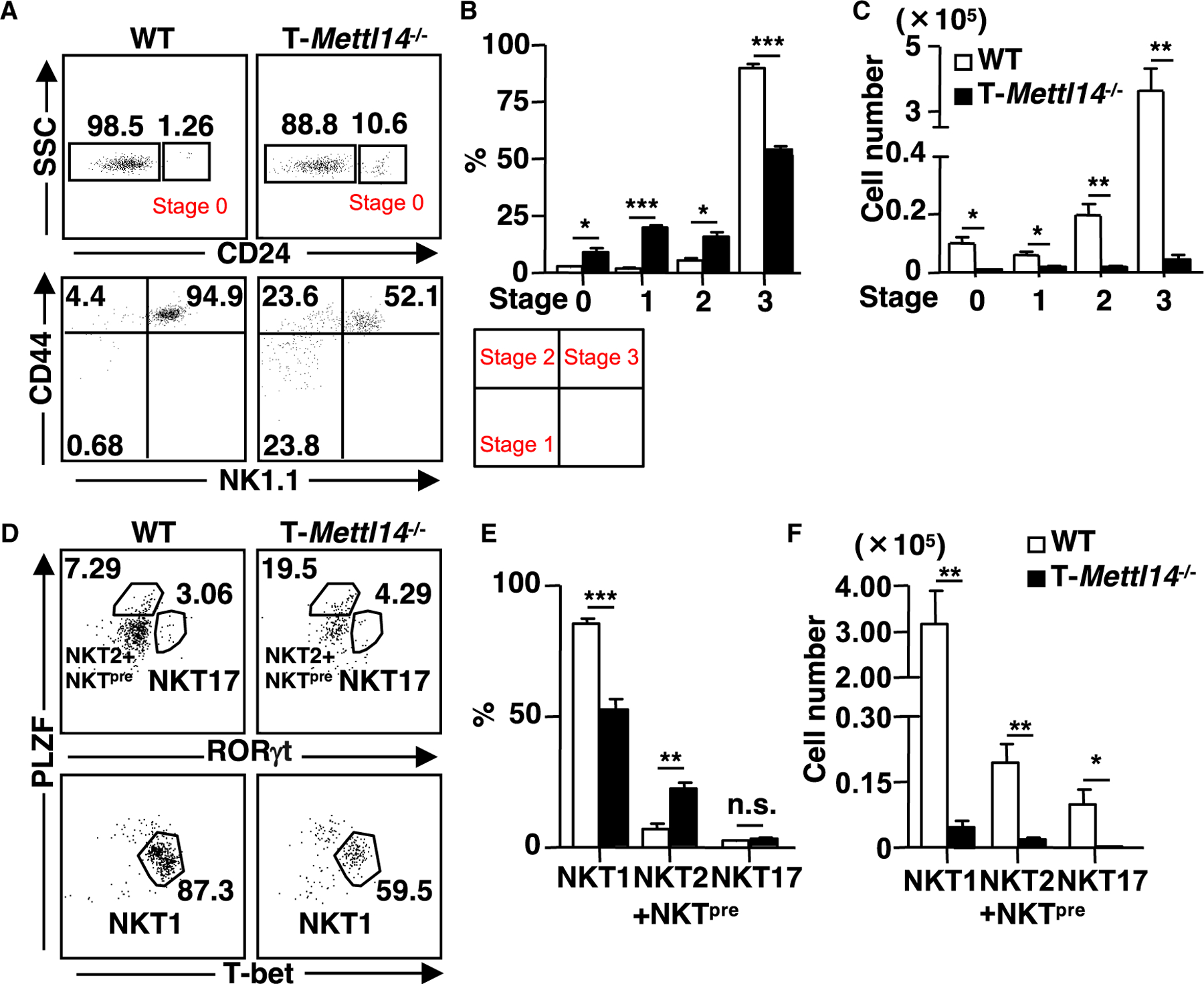
(A) Representative staining of iNKT cells (CD69+CD1d/PBS57 tetramer+) at various developmental stages in the thymus of WT and T-Mettl14−/− mice.
(B and C) Quantification of percentages and cell numbers of stages 0, 1, 2, and 3 iNKT cells in WT and T-Mettl14−/− mice (n = 3–4).
(D) Intracellular staining of PLZF, T-bet, and RORγt in thymic iNKT cells of WT and T-Mettl14−/− mice.
(E and F) Quantification of percentages and cell numbers of NKT1, NKT2+NKTpre, and NKT17 subsets (n = 6). SEM is shown. *p < 0.05, **p < 0.01, ***p < 0.001.