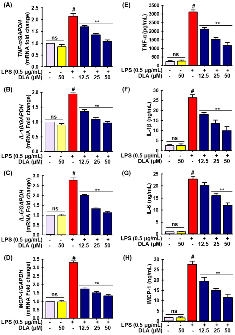
Figure 3.
DLA impact on the production of proinflammatory cytokines and chemokine. A 96-well plate was seeded with cells (2 × 10⁵ cells/mL) and incubated for 24 h. Following this, the cells were exposed to DLA (12.5–50 µM) or vehicle alone for 20 h and/or for 3, then to LPS (500 ng/mL), and finally they were incubated for 6 h. DLA impact on LPS-induced RAW 264.7 cells mRNA expression of tumor necrosis factor-α (TNF-α), interleukin-1β (IL-1β), interleukin-6 (IL-6), and monocyte chemoattractant protein-1 (MCP-1) (A–D) and protein expression (E–H). # p < 0.05 vs. treatment with vehicle control; ** p < 0.05 vs. treatment with LPS alone. ns: non-significant vs. treatment with vehicle control.

