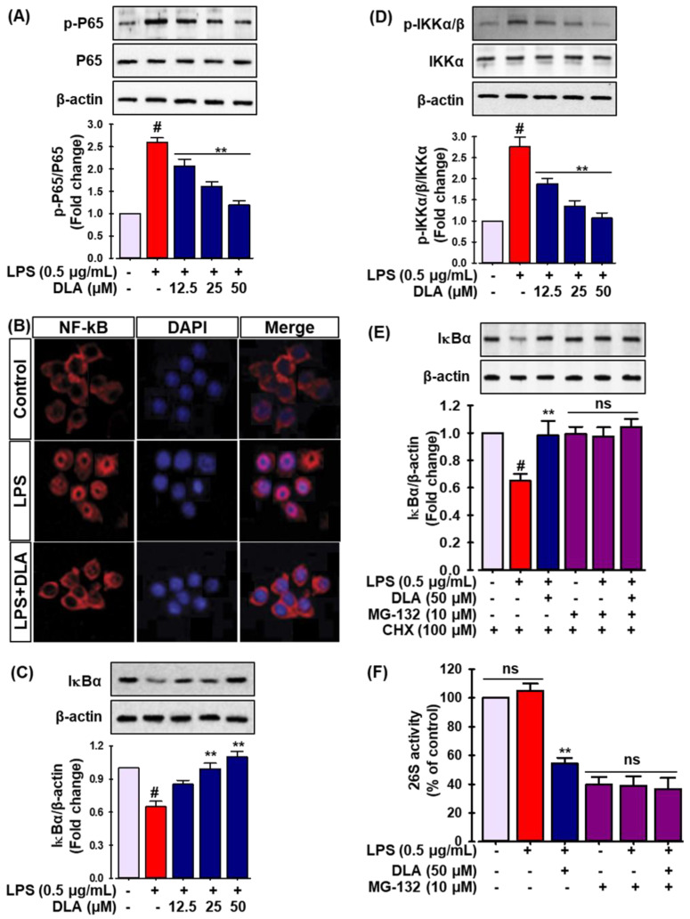
Figure 4.
Effect of DLA on the nuclear factor kappa-light-chain-enhancer of activated B cells (NF-κB) signaling pathway. The cells were pretreated with DLA (12.5, 25, and 50 μM) for 12 h before being exposed to LPS (500 ng/mL) for 1 h. Immunoblot analysis was performed to evaluate the protein levels of p-p65 and p65 (A); # p < 0.05 vs. vehicle-treated control; ** p < 0.05 vs. treatment with LPS alone. The cells were fixed for immunofluorescent labeling for p65. Red represents p65; blue represents DAPI staining of the nuclei (B). The protein levels of inhibitor protein κBα (IκBα) (C), IκB kinase p-IKKα/β, and IKKα/β (D) were quantified by Western blot; # p < 0.05 vs. vehicle-treated control; ** p < 0.05 vs. treatment with LPS alone; Cells were treated with 500 ng/mL of LPS and 10 μM MG-132 for 30 min. Following cell harvesting, the protein level of IκB was assessed by Western blot analysis, and the relative change was determined (E), the cells were pre-treated with 50 μM DLA for 12 h and then incubated with 10 μM MG-132 for 1 h; After 30 min of treatment with 500 ng/mL of LPS, the proteasome activity in the cell lysates was determined (F); # p < 0.05 vs. vehicle-treated control; ** p < 0.05 vs. treatment with LPS alone, ns: non-significant vs. treatment with MG-132 alone.

