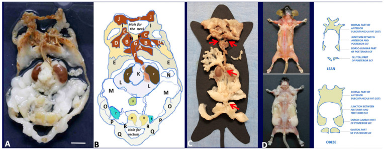
Figure 1.
Gross anatomy of mouse adipose organ in different experimental conditions. (A): Anatomical preparation of the adipose organ as a unitary structure (adult, male C57BL/6 mouse, normoweight). Subcutaneous-visceral continuities (SVCs) are well visible and indicated in the diagram legend. Preputial glands and kidneys are left to facilitate anatomical orientation. White (WAT) and brown (BAT) adipose tissues are recognizable by their specific colors. (B): Legend: A: interscapular BAT (subcutaneous, SC), B: subscapular BAT (SC), C: supraclavicular BAT (SC), D: axillary BAT (SC), E: axillo-thoracic WAT (SC), F and G: periaortic mediastinal BAT (Visceral, V), H: SVC, I: cervical subcutaneous WAT (SC), J: cervical subcutaneous BAT (SC), K: mesenteric WAT (V), L: perirenal-retroperitoneal WAT-BAT (V), M: epididymal WAT (V), N: omental WAT (V), O: dorso-lumbar WAT (SC), P: inguinal WAT (SC), Q: gluteal WAT (SC). A + B + C + D + E + I + J: anterior subcutaneous region of adipose organ. O + P + Q: posterior subcutaneous region of the adipose organ. F + G + K + L + M + N: visceral region of the adipose organ. H and R: SVCs. r: kidney, s: urinary bladder, u: preputial glands. (C): Anatomical preparation of the adipose organ (adult, female C57BL/6 mouse, normoweight) as a unitary structure laying on a template. Red arrows point to SVCs (H and R in (B)) and mesenteric-abdominopelvic (inter-visceral) connection. (D): Dorsal view of adult C57BL6 female mice after skin removal. Anterior and posterior subcutaneous parts of the adipose organ are visible in both normoweight (upper panel) and obese animals. Bar: 8 mm in (A,B), 13 mm in (C) and 40 mm in (D).

