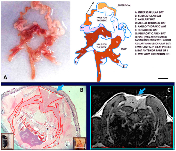
Figure 2.
The fat neckerchief of mouse Adipose Organ. (A): Anatomical preparation of fat neckerchief displaying its continuity with peri-aortic mediastinal fat (adult C57BL/6 at room temperature). The superficial white fat of the neckerchief is separated and folded up as indicated by the arrow in the diagram. (B): Histology (newborn, C57BL/6) and axial T1-weighted MRI image (adult, C57BL/6) (in (C)) of the interscapular region, show the continuity between interscapular fat depot with the axillary fat, outlined by the red line surrounding brown fat in histology. Further evidence is shown by serial sections in histology and MRI (Supplementary Figure S7). The interscapular region (indicated by light blue arrows) also shows that brown (identified by a hypointense area) and white adipose tissue (hyperintense area) are contained in the same depot as also evident in anatomical preparations and histology. Insets in (B) show the plane of the section in the whole animal (left) and in its sagittal section (right). Bar: 10 mm in (A,B), and 1.5 mm in (C).

