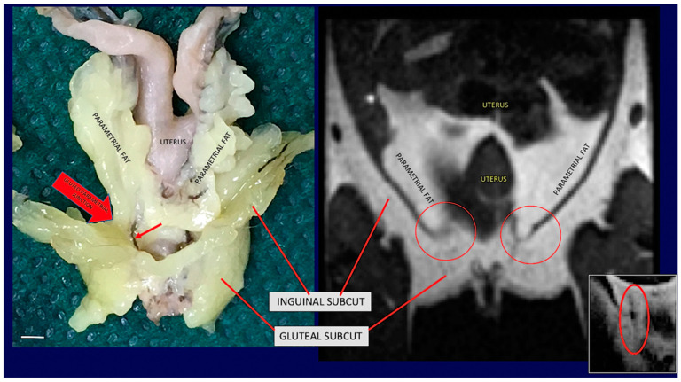
Figure 3.
Gross anatomy and MRI of subcuteous-visceral continuity in the lower part of mouse Adipose Organ. (Left): Anatomical preparation of an adult C57BL/6 mouse showing subcutaneous-visceral continuity between parametrial and gluteal fat. (Right): coronal T1-weighted MRI of the same areas shown in the left panel of the same animal in vivo. Anatomical continuity is indicated in the visceral (parametrial) and subcutaneous (inguinal) fat by arrow and red circles. A small red arrow on the left panel shows a vessel in the junction. A similar structure was also detected by MRI (red circled area in small, squared panel on the right). Bar 4.5 mm in both panels.

