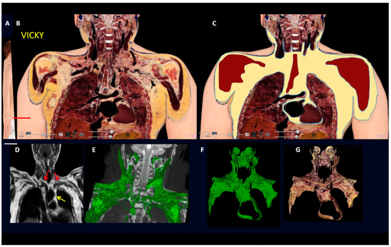
Figure 4.
Gross anatomy and MRI of subcutaneous-visceral continuity in human Adipose Organ. (A): Oblique frontal section of a digital cadaver (Vicky). (B): Supraclavicular-mediastinal continuity of the section shown in (A). (C): The same section shown in (B), with the fat area highlighted in yellow. (D): Routine MRI in an adult male. Red arrows indicate subcutaneous-visceral fat connection (SVC) and the yellow arrow indicates the mediastinal fat. (E): 3D rendering (green) of the SVC in a 10-year-old boy. (F,G): 3D reconstruction of the fat present in the supraclavicular-mediastinal continuity shown in (B,C). Fat area is highlighted in three planes (axial, coronal, and sagittal); in F fat is in green for comparison with (E); in (G) fat is shown in its original colors. Bar: 60 mm in (B,C), 45 mm in (F,G).

