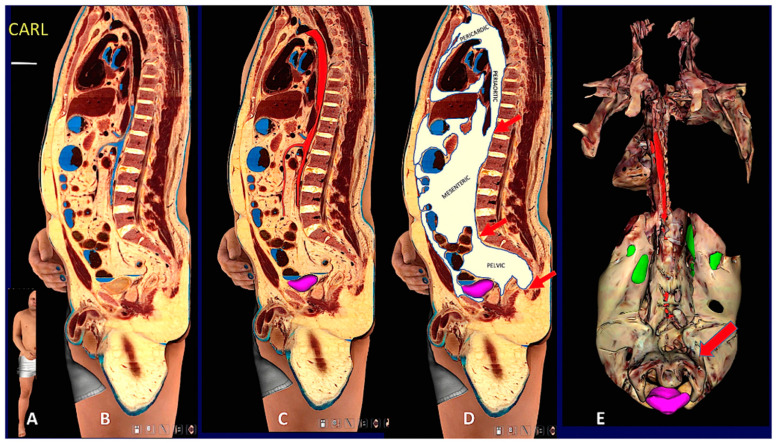
Figure 6.
Digital cadaver section and 3D reconstruction showing upper and lower subcutaneous visceral continuity in the Adipose Organ. (A): Sagittal oblique section of the digital cadaver, Carl. (B–D): Mediastinal-abdomino-pelvic and gluteal subcutaneous-visceral connection (SVC). (B): original section, (C): aorta and upper mesenteric artery in red, (D): fat highlighted in white. Arrows in (D) point to mediastinic-abdominal (upper), abdomino-pelvic (middle) and gluteal-pelvic (lower) connections. Gluteal-pelvic SVC is also shown in Supplementary Figure S16. Panel (E) shows a 3D reconstruction of the whole visceral fat. Dorsal view in which the continuity between retroperitoneal and pelvic fat surrounding the urinary bladder is more evident, indicated by the arrow. Green: kidneys (parts of dorsal surface not covered by retroperitoneal fat), violet: urinary bladder. Arrows point to periaortic fat in the mediastinic-abdominal connection. See also Suppl movie. Bar: in (B–D) 60 mm, in (E) 120 mm.

