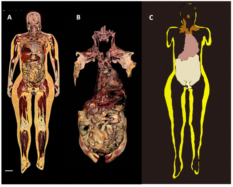
Figure 7.
2D and 3D reconstructions of human Adipose Organ. Digital cadaver Vicky. (A): Representative images showing the whole adipose organ in a coronal section, (B): the 3D reconstruction of isolated visceral fat in connection with the supraclavicular-axillary part of the adipose organ, and (C): the visceral (brown, beige, and pale white) and subcutaneous (yellow) parts of the adipose organ (still image from S Movie 1). Bar: 65 mm in (A), 35 mm in (B) and 75 mm in (C).

