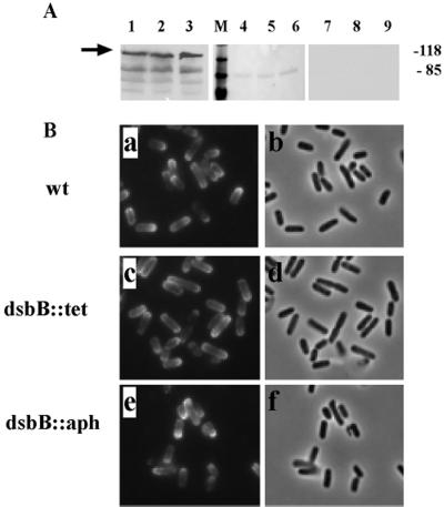
FIG. 4.
Effects of the dsbB mutation on the surface presentation and unipolar targeting of IcsA. (A) Expression and surface cleavage of IcsA. Lanes 1 to 3, Western blot analysis of total cellular protein extracts (arrow indicates IcsA); lanes 4 to 9, supernatant proteins on nitrocellulose, stained with Ponceau S, showing presence of protein (lanes 4 to 6), or probed by Western blot for IcsA, showing lack of secreted and cleaved IcsA (lanes 7 to 9). Lanes 1, 4, and 7, 2457T (wild type [wt]); lanes 2, 5, and 8, LDB143 (dsbB1); lanes 3, 6, and 9, LDB660 (dsb::aph). Apparent molecular masses of standard proteins run in parallel are indicated (in kilodaltons). (B) Localization of IcsA on the bacterial surface. Indirect immunoflourescence (a, c, and e) and corresponding fields by phase microscopy (b, d, and f). (a and b) 2457T; (c and d) LDB143; (e and f) LDB660.

