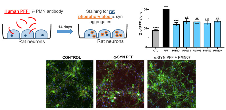
Figure 11.
mAb inhibition of the recruitment of endogenous rat α-syn into phosphorylated aggregates. Cultures of primary rat hippocampal neurons were exposed to sonicated human PFF (1 μg/mL) without or with mAbs (0.05 μM, except for PMN09 at 0.25 μM). CTL = neurons incubated with vehicle alone. Cultures were stained 14 days later for neuronal marker MAP2 (green), aggregates of phosphorylated rat α-syn (red) and cell nuclei (blue). Results are expressed as a percentage of the phosphorylated rat α-syn staining area with PFF alone and show the mean ± SEM of 6 replicate cultures. A global analysis of the data was performed using one-way ANOVA followed by Dunnett’s multiple comparisons test. ** p ≤ 0.01, *** p ≤ 0.001, **** p ≤ 0.0001 vs. PFF.

