Figure 6.
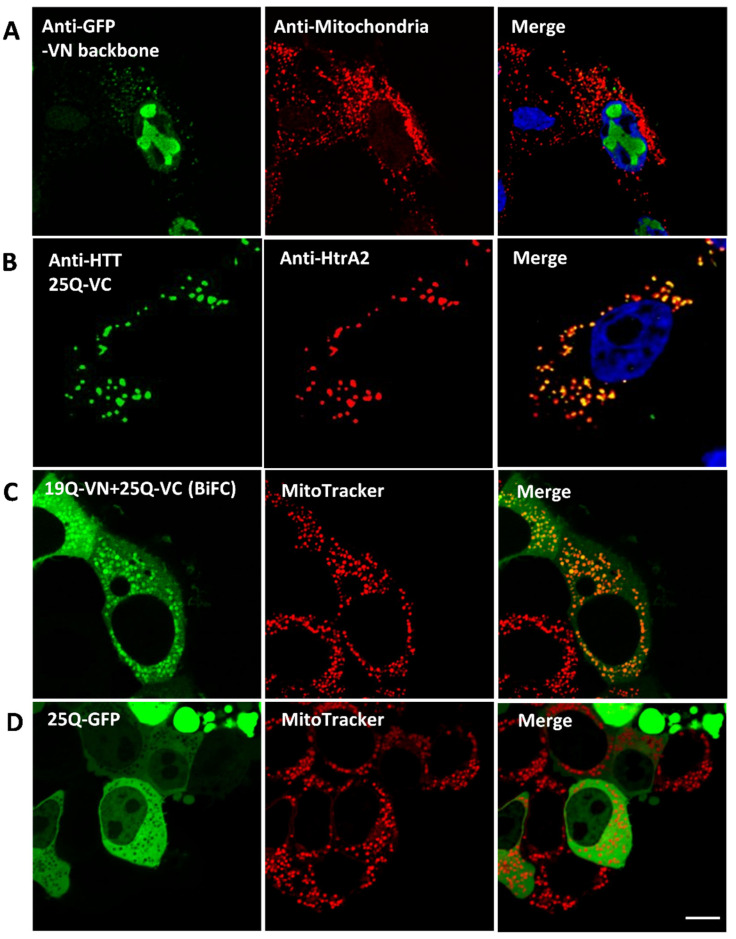
Fluorescence tag effect on wild type HTT subcellular localisation in fixed HEK293T cells transfected with various constructs for 48 h. (A) N-terminal half of Venus (VN) localisation in dual immunolabelled HEK293T cells. Left panel: anti-GFP (ab6556) (Alexa Fluor 555). Middle panel: anti-mitochondria antibody (MAB1273) (Alexa Flour 647). Right panel: merge of VN and mitochondrial signals, nuclei stained with Hoechst 33342. VN localises mostly in the nucleus, with minor punctate staining in the cytosol which co-localises with mitochondria. (B) Cells expressing 25Q-VC were double immunolabelled as follows: Left panel: anti-HTT (mEM48) antibody (MAB5374) (Alexa Fluor 647). Middle panel: anti-HtrA2/Omi antibody (AF1458) (Alexa Fluor 555). Right panel: merge of HTT and HtrA2 signals; nuclei were stained with Hoechst 33342. 25Q-VC co-localises with mitochondria. (C,D) Cells were seeded in ibiTreat dishes and co-transfected for 48 h with 19Q-VN and 25Q-VC (C) or 25Q-GFP (D). Live cells were stained with MitoTracker Red CMXRox (M-7512) prior to confocal examination. Acquired images were deconvolved. (C) Left panel: BiFC signal 19Q-VN and 25Q-VC. Middle panel: MitoTracker signal. Right panel: merge of BiFC and MitoTracker signals. BiFC signal of WT HTT is mitochondrial (indicated by the co-localisation with MitoTracker) as well as cytosolic. (D) Left panel: GFP signal showing the fused 25Q localisation. Middle panel: MitoTracker signal. Right panel: merge of the 25Q-GFP and MitoTracker signals. Scale bar = 8 µm. 25Q-GFP is expressed in the cytosol, with complete exclusion of mitochondrial localisation.

