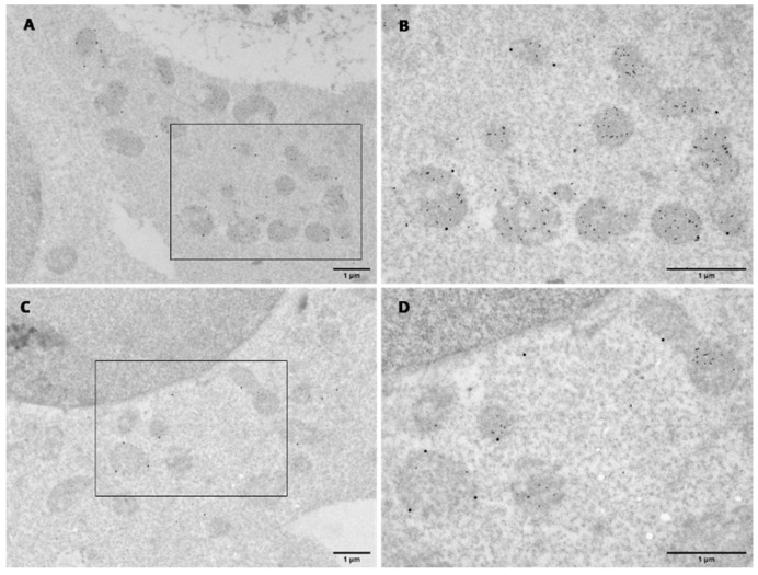
Figure 7.
Electron micrographs of dual immunogold labelling in transfected HEK293T cells for 48 h. Cells were co-transfected and co-probed with anti-KMO antibody (10698-1-AP) and anti-HTT (mEM48) antibody (MAB5374), followed by 30 and 15 nm gold conjugate secondary antibodies, respectively. (A,C) Overview of dual labelling of flKMO-CC and 19Q-VN, respectively. Scale bar = 1 µm. (B,D) Zoomed view of the regions indicated by the black box in (A) and (C), respectively. Mitochondria are all intensely labelled with HTT (15 nm particles), and some particles are seen in the cytoplasm. flKMO-CC labelling is seen on the outer membrane of some HTT-labelled mitochondria (30 nm particles). Scale bar = 1 µm.

