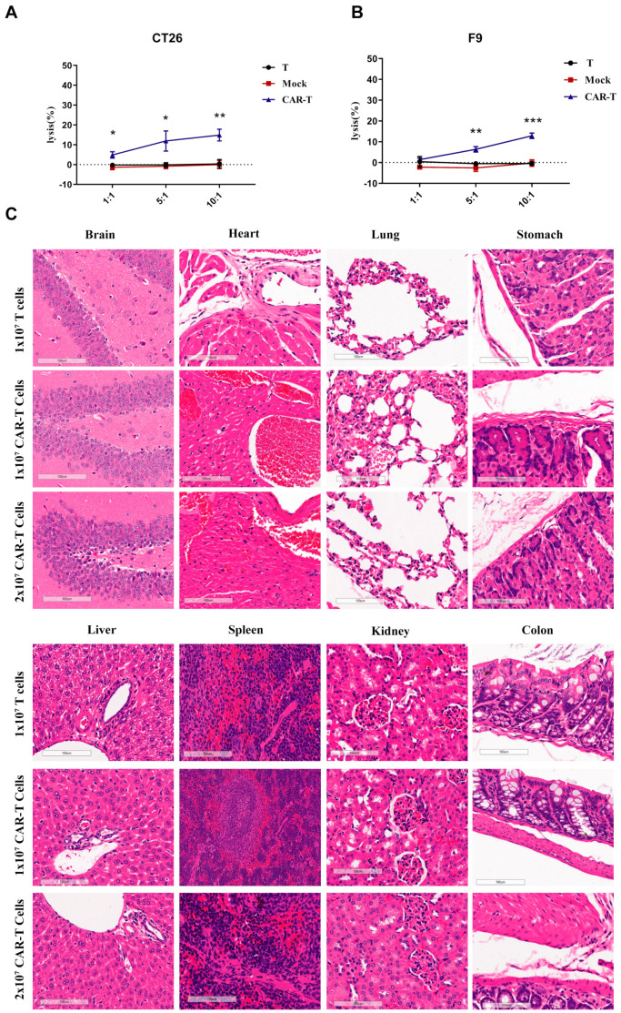
Figure 5.
Histopathological analysis of murine organ tissues by hematoxylin and eosin staining. (A,B) Human EDB-targeted CAR-T-cells incubated with mouse tumor F9 or CT26 cells for 24 h. Cell lysis was determined using an LDH assay. (C) NCG mice were treated with 1 × 10⁷ T-cells and 1 × 10⁷ or 2 × 10⁷ EDB CAR-T-cells and sacrificed on Day 21 following T-cell infusion. Different tissues were harvested, formalin-fixed, paraffin-embedded, and stained with H&E. Representative photomicrographs are shown. The images were taken through a Leica Aperio VERSA 8 slice scanner under 20× magnification. Each scale bar represents 100 μm. No obvious pathological changes were found in any tissues, and there were no significant differences between all groups. Datapoints reflect the mean ± SD of triplicates (* p < 0.05; ** p < 0.01; *** p < 0.001; two-tailed Student’s t-test).

