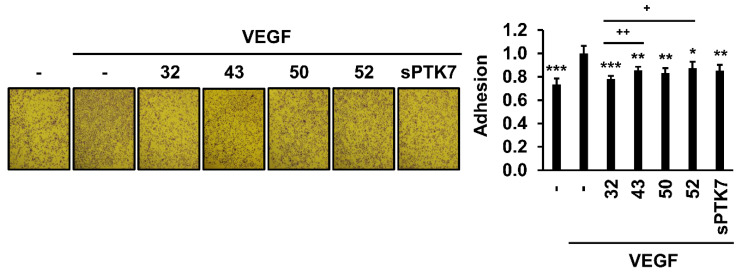
Figure 2.
Effect of anti-PTK7 mAbs on the adhesion of HUVECs. Adhesion of HUVECs was assessed for 1 h in the presence of anti-PTK7 mAbs (mAb-32, mAb-43, mAb-50, and mAb-52; 10 μg/mL) or sPTK7 (4 μg/mL) after plating the cells on a 96-well plate whose wells were pre-coated with 0.1% gelatin. The number of adhered cells was measured after crystal violet staining. Representative images are shown. Magnification = ×40. “–”: no treatment with anti-PTK7 mAb or sPTK7. The graph shows the number of adhered cells in the mAb-treated groups relative to that in the VEGF-alone group (n = 5). Data represent mean ± standard deviation. * p < 0.05, ** p < 0.01, and *** p < 0.001 vs. VEGF-alone control. + p < 0.05 and ++ p < 0.01 vs. mAb-32-treated group. The absence of the p value between antibody-treated groups indicates statistical insignificance.

