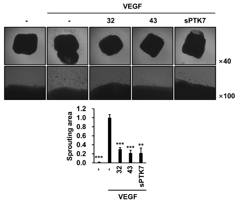
Figure 7.
Effect of PTK7 mAbs on VEGF-induced angiogenesis ex vivo. Mouse aortic ring assay was performed to evaluate angiogenesis ex vivo. Mouse aortas were placed on solidified growth-factor-reduced Matrigel and incubated in M199 medium with 1% FBS and with or without mouse VEGF (20 ng/mL) in the presence of mAb-32 or mAb-43 (10 μg/mL) or sPTK7 (4 μg/mL) for 12 days. Outgrowth of endothelial cells from aortas was observed under a phase-contrast microscope (magnification = ×40 and ×100). “–”: no treatment with anti-PTK7 mAb or sPTK7. The graph shows the sprouting area from the aorta in the mAb-treated and sPTK7-treated groups relative to that in the VEGF-alone group (n = 3). Data are represented as mean ± standard deviation. ** p < 0.01 and *** p < 0.001 vs. VEGF-alone control. The absence of the p value between antibody-treated groups indicates statistical insignificance.

