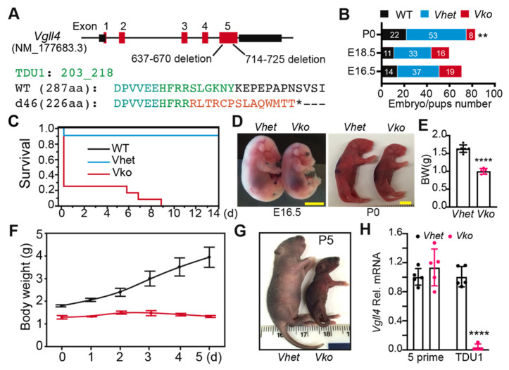
Figure 2.
VGLL4 deletion resulted in perinatal lethality: (A) Vgll4 gene structure showing Vgll4-mutant allele. Green and red letters indicate wild-type and d46 mutant amino acid sequences, respectively; * indicates a premature stop codon; (B) distribution of genotypes at E16.5, E18.5, and P0. Embryo/pup numbers for each genotype are displayed in the bar graph. **, Mendelian ratio chi-squared test, p < 0.01; (C) survival curve of Vgll4 d46/+ (Vhet) and Vgll4 d46/d46 (Vko) pups. Postnatal day 0 (P0) was designated as the date of delivery. For each group, N = 12; (D) gross morphology of embryos and mouse pups at indicated ages. Scale bar: 5 mm; (E) body weight at E18.5; (F) mouse pups’ body weight gain in the first 5 days after delivery. N = 3; (G) gross morphology of mouse pups at P5; (H) cardiac Vgll4 mRNA expression. E13.5 hearts were collected for mRNA isolation and qRT-PCR measurement. TDU1 domain of Vgll4 was not detectable in the Vgll4 mutant transcripts. (E,H), Student’s t-test: **** p < 0.0001. N = 5–6.

