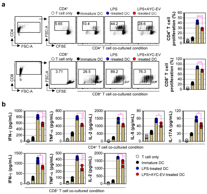
Figure 5.
The capacity of AYC-EVs to regulate LPS-treated DCs-mediated T cell proliferation and activation. (a,b) splenic CD4+ and CD8+ T cells sorted from BALB/c mice were stained with CellTrace (CFSE), and CFSE-labeled T cells were co-cultured with DCs treated with PBS (immature DC), LPS (100 ng/mL), or LPS with AYC-EVs (20 μg/mL) in presence of anti-CD3 (1 μg/mL) and anti-CD28 (1 μg/mL). Following incubation for 48 h, cells and supernatants were harvested. (a) The cells were stained with anti-CD4 and anti-CD8 antibodies. Levels of T cell proliferation (CD4+CFSE- and CD8+CFSE- cells) were evaluated using flow cytometry. (b) Cytokine production in CD4+ and CD8+ T cells was measured in culture supernatants using cytokine-specific ELISA kits. All bar graphs display mean ± SD (n = 3 samples). All experiments were repeated three times with similar results. * p < 0.05, ** p < 0.01, *** p < 0.001.

