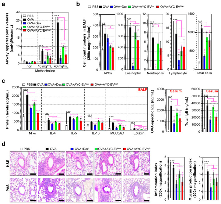
Figure 6.
Effects of AYC-EV administration on airway hyper-responsiveness, cytokine production, and histological changes in an OVA-induced asthma model. (a) Airway hyper-responsiveness was evaluated using Flexivent 24 h after the last OVA challenge in an OVA-induced asthma model. (b) The inflammatory cells (eosinophils, APCs, neutrophils, and lymphocytes) in BALF were deposited on slides and stained with Diff-Quik stain reagent and counted in a double-blind manner in 3 areas for each slide. (c) The levels of TNF-α, IL-4, IL-5, IL-13, MUC5AC, and eotaxin in the BALF and the OVA-specific IgE and total IgE in the serum were determined using ELISA. (d) Histopathological analysis of airway inflammation and mucus production was performed in the lung tissues using H&E and PAS staining. Scale bars, 100 μm. PBS: OVA: group administrated with PBS; OVA: group administrated with OVA; OVA+Dex: group administrated with OVA and dexamethasone (3 mg/kg); OVA+AYC-EVlow: group administrated with OVA and 4 mg/kg AYC-EVs; and OVA+AYC-EVhigh: group administrated with OVA and 8 mg/k1000g AYC-EVs. Values are presented as means ± SD (n = 7 mice). * p < 0.05, ** p < 0.01, *** p < 0.001. APCs: antigen-presenting cells.

