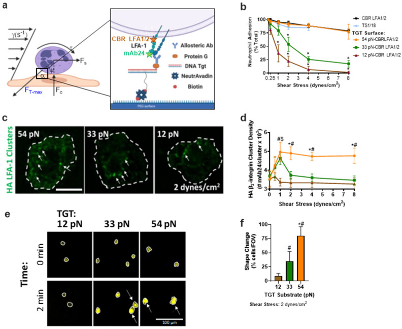
Figure 1.
Neutrophils’ adhesion strengthening depends upon the level of bond tension acting on HAβ2-integrin. (a) Schematic of neutrophils sheared in microfluidic channels bound to the glass coverslip via allosteric antibody, CBR LFA1/2 (binding epitope shown in orange) linked to one side of a TGT that is annealed to the substrate on glass coverslips. High-affinity β2-integrin was reported on using mAb24 (binding epitope shown by green star) (b) Neutrophils binding to each TGT bearing CBR LFA1/2 substrate quantified as the percentage of cells that remained bound after incrementally increasing shear stress, as defined on the x-axis. Arrested neutrophil percent is reported as mean +/− SEM over 5 fields of view with >10 cells per experiment (n = 3, * compared to CBR LFA1/2, p < 0.01). (c) AF488 mAb24 clusters (green) of bound neutrophils sheared over CBR LFA1/2:TGT substrate at (1 dynes/cm2) measured via TIRF; scale bar depicts 5 µm. (d) HA β2-integrin cluster density measured via TIRF. Graphs plot HA β2-integrin clustering mean +/− SEM for 5 fields of view for each experiment; scale bar shows 10 µm (n = 3, * 54pN and 33pN, # 54pN and 12pN, $ 33pN and 12pN, p < 0.01). (e) Neutrophils with Ca2+ reporter Rhod-2 sheared over TGT/HA-inducing β2-integrin substrate for 2 min at 2 dynes/cm2. White circles show area and arrows identify cells that have undergone shape change compared to time 0; scale bar shows 100 µm. (f) Quantification of the percentage of neutrophils that change shape after shearing for 2 min over TGT/HA-inducing β2-integrin substrate. Graphs plot percent shape change mean +/− SEM for 5 fields of view for each experiment (n = 3, * 54pN and 33pN, # 54pN/33pN and 12pN, p < 0.01, n.s.: not significance).

