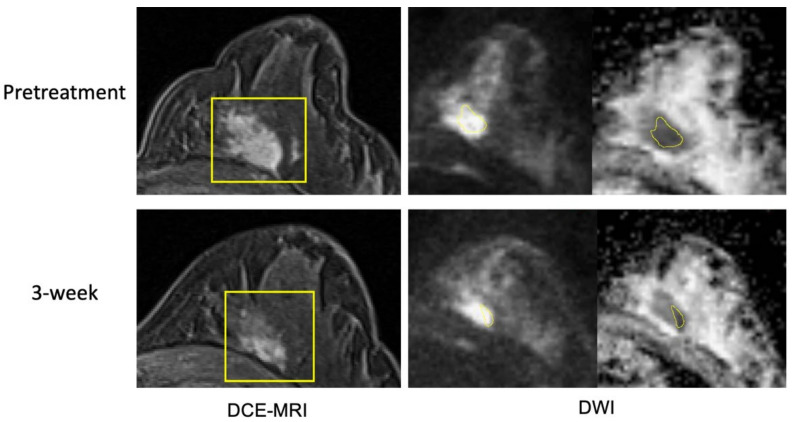
Figure 5.
Example case 2 without a pathologic complete response. The patient was treated by paclitaxel + pembrolizumab followed by cyclophosphamide. Representative MR images are shown from pretreatment (top row) and at 3-week (bottom row). Images from DCE-MRI were acquired 119 s after the contrast injection. Images from DWI are shown in a pair of original DWI (b = 800 s/mm2) and ADC map. ROIs are shown in yellow (rectangular box in DCE and hand-drawn in DWI). Images from pretreatment and at 3-week may seem different by visual comparison due to different positioning in the MRI scanner. Breast tissue in DCE-MRI suggests that they are from similar slice location.

