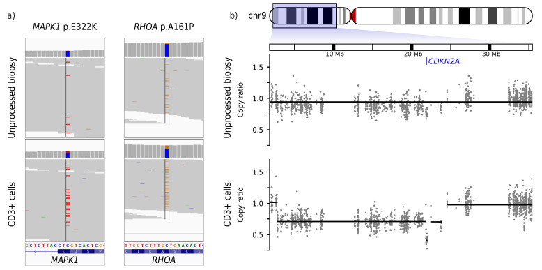
Figure 2.
Somatic variant calling in an early-stage MF skin biopsy without (top) and with (bottom) enrichment of tumor cells. One skin lesion of a patient with tumor stage 1 MF was biopsied. One half (unprocessed biopsy) was used without further steps. The other half of the material was used for enrichment of CD3-positive cells (CD3+ cells). Afterwards, both halves were used for DNA isolation, WXS, and calling of somatic SNVs and CNVs. (a) Raw data for the somatic SNVs MAPK1 p.E322K and RHOA p.A161P where colored strips indicate variant reads diverging from the genome reference. (b) Raw and processed data for a deletion on chr9 leading to homozygous loss of CDKN2A. The gray dots show the copy-ratio (denoised and normalized read depth of individual exons) while the black lines depict the automatic CNV call. A copy-ratio of 1 indicates a neutral copy-number of 2 on autosomes. The genomic position of CDKN2A is marked in blue.

