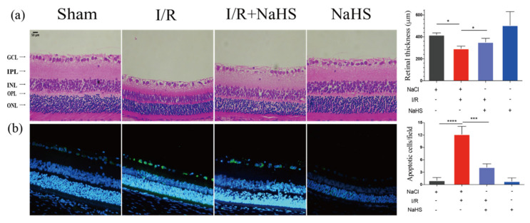
Figure 2.
NaHS alleviated I/R-induced retinal neurodegeneration. (a) Images of hematoxylin and eosin staining and overall thickness of retinae quantitative analysis. NaHS improved the retinal thinning induced by cerebral I/R. (b) Representative images of TUNEL staining and quantitative analysis of TUNEL+ (apoptotic, green) cells. NaHS decreased apoptotic cells in GCL. Scale bar = 50 μm. Data are presented as the means ± SD, n = 5, * p < 0.05, *** p< 0.001, **** p< 0.0001. Ganglion cell layer—GCL; interplexiform layer—IPL; Inner Nuclear Layer—INL; outer plexiform layer—OPL; outer nuclear layer—ONL.

