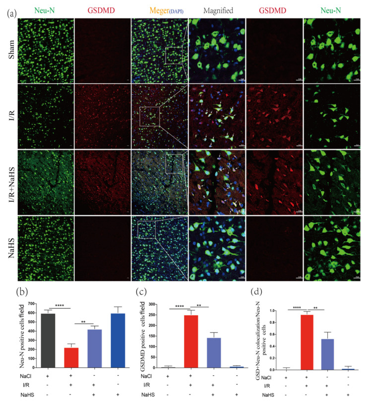
Figure 5.
NaHS inhibited I/R-induced neuron pyroptosis in the rat brain cortex. (a) Immunofluorescence staining of Neu-N and GSDMD in the peri-infarct region. Scale bar = 50 μm. Scale bar = 25 μm in magnified images. Arrows indicate Neu-N and GSDMD double-positive cells. (b) Analysis of Neu-N Positive cells in the brain cortex (n = 3). (c) Analysis of GSDMD-positive cells in the brain cortex (n = 3). (d) Analysis of the ratio of the Neu-N and GSDMD double-positive cells to the Neu-N positive cells in the brain cortex (n = 4). Data are represented as means ± SD. ** p < 0.01, **** p < 0.0001.

