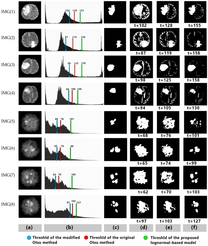
Figure 4.
Selected samples, IMGs (1), (2), (3), and (4) are MRI brain tumor images, IMGs (5), (6), (7), and (8) are simulated images; (a) are original images, (b) corresponding histograms, (c) ground truths, (d) segmentation results using the modified Otsu method [3], (e) segmentation results using the original Otsu, and (f) segmentation results using the proposed lognormal-based model.

