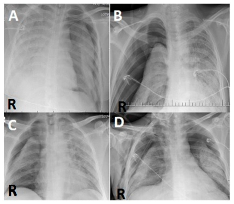
Figure 2.
(A) In a posterior–anterior lung roentgenogram, the right lung is almost completely radiopaque, while PNX is seen on the left side. (B,C) In a posterior–anterior lung roentgenogram, the left lung is almost completely radiopaque, while PNX is seen on the right side. (D) Bilateral PNX is observed in aposterior–anterior lung roentgenogram (R: right).

