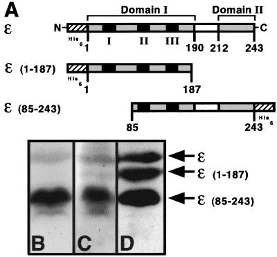
FIG. 2.
(A) Structures of the ɛ derivatives used in this study. Proposed domains of ɛ are indicated (33). Amino acid residues are indicated, as are the three exonuclease motifs (black boxes), the proposed N- and C-terminal domains (gray boxes) and Q linker (white boxes), and the hexa-histidine tags (cross-hatched boxes). Note that the figures are not to scale. Polyvinylidene difluoride membranes (Millipore) containing the indicated ɛ derivatives (1 μg each) after fractionation by sodium dodecyl sulfate-polyacrylamide gel electrophoresis were processed either as a Far Western blot (31) by probing with 32P-labeled UmuD (B) or UmuD′ (C) or as a standard Western blot (D) using antipolyhistidine tag antibodies as described elsewhere (23, 31). The relative position of each ɛ derivative is indicated.

